 Fenwickgoods
Analyze the relationship between microscopic Z-disc tearing and actual hypertrophy. You will see why severe soreness often impairs subsequent training adaptations.
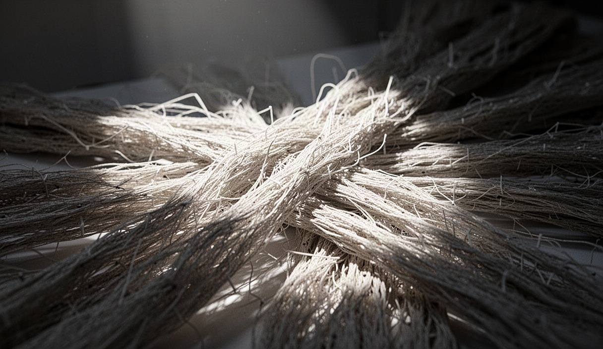
For decades, lifters and coaches treated severe post-exercise muscular soreness as the premier badge of an effective workout. The prevailing hypothesis stated that resistance training must tear down muscle fibers to initiate an adaptive rebuild, implying that greater microscopic tearing equates to a larger muscular adaptation. In strength science, this phenomenon is categorized as exercise-induced muscle damage. It presents visibly as torn sarcomeres, disrupted cell membranes, and delayed-onset soreness that peaks two days later.
Recent advances in muscle biology and mechanotransduction have reshaped this narrative. Mechanical tension remains the primary trigger for myofibrillar accretion, while muscle damage represents an inevitable, often counterproductive byproduct of high-force contractions at long muscle lengths. Dissecting the precise biological pathways of structural damage reveals how the body resolves tissue injury and why chasing damage directly often stalls muscular development instead of accelerating it.
The Mechanics of Micro-trauma
Micro-trauma occurs primarily during the eccentric phase of a muscular contraction, where muscle fibers are forcibly lengthened while attempting to shorten. Under the sarcomere popping hypothesis, individual sarcomeres along a myofibril do not stretch uniformly. When a muscle fiber is stretched past its optimal filament overlap, the weakest sarcomeres yield first, extending beyond mechanical stability. This non-uniform elongation creates high shear stress across adjacent structures, causing physical disruption to the internal lattice of the cell.
The earliest morphological indicator of this micro-trauma is Z-line streaming. The Z-disc, which anchors the thin actin filaments within each sarcomere, loses its rigid, parallel alignment and smears across the intracellular space. This structural failure degrades intermediate filaments, specifically desmin, which binds adjacent myofibrils together, and dystrophin, which links the internal cytoskeleton to the extracellular matrix. Once these structural scaffolds buckle, the membrane integrity of the sarcolemma is compromised.
A compromised sarcolemma permits an uncontrolled influx of extracellular calcium into the cytosol. Under basal conditions, intramuscular calcium is tightly regulated within the sarcoplasmic reticulum. The sudden cytosolic calcium surge activates calpains, which are calcium-dependent neutral proteases. Calpains immediately degrade structural proteins, including titin, nebulin, and the remaining desmin lattice, converting localized mechanical strain into rapid enzymatic auto-digestion within hours of the training stimulus.
| Structural Level | Primary Component Damaged | Functional Consequence |
|---|---|---|
| Sarcomere | Z-line alignment and actin-myosin cross-bridges | Immediate loss of active force production |
| Cytoskeleton | Desmin and titin proteins | Lateral force transmission failure between myofibrils |
| Sarcolemma | Phospholipid bilayer and dystrophin complex | Calcium ion leakage, protease activation, cellular edema |
| Extracellular Matrix | Collagen network and lamina densa | Altered stiffness and increased sensory nerve sensitivity |
Satellite Cell Activation Dynamics
Once micro-trauma occurs, the muscle fiber depends on a dedicated population of myogenic stem cells, known as satellite cells, located between the basal lamina and the sarcolemma. In healthy, resting tissue, satellite cells remain quiescent, expressing the paired-box transcription factor Pax7. When mechanical trauma breaches the sarcolemma, local inflammatory signaling molecules, such as hepatocyte growth factor and basic fibroblast growth factor, dislodge Pax7-positive cells from their quiescent niche, initiating rapid proliferation.
Following proliferation, these activated cells transition into myoblasts by up-regulating regulatory factors including MyoD and Myf5. The myoblasts then proliferate across the injured section of the fiber before down-regulating Pax7 and up-regulating myogenin, signaling the terminal differentiation phase. These differentiated cells can either fuse directly with an injured, existing fiber to donate their nuclei, or fuse with one another to form an entirely new, embryonic myosin heavy chain-expressing myotube to replace a fully necrotic fiber segment.
The critical biological distinction lies between regenerative repair and net hypertrophy. When satellite cells fuse with an extensively damaged muscle segment, their nuclear donation primarily serves to re-establish normal protein turnover rates across a disrupted myonuclear domain, which generally covers 2000 to 2200 square micrometers of cytoplasm per nucleus. If the muscle damage is minimal to moderate, mechanical tension can stimulate satellite cell donation without extensive structural necrosis, allowing the added myonuclei to support net increases in cross-sectional area rather than simply restoring basal cellular architecture.
The Energy Cost of Tissue Repair
Repairing damaged muscle is biologically expensive. When fibers tear, the local environment releases intracellular contents into the bloodstream, triggering an immediate inflammatory cascade. Neutrophils arrive at the injury site within 2 to 6 hours, followed by pro-inflammatory M1 macrophages. These immune cells perform phagocytosis, engulfing and enzymatically dismantling necrotic protein fragments via reactive oxygen species and lysosomal proteases. This clearing of cellular debris consumes large pools of adenosine triphosphate (ATP) before any structural rebuilding can begin.
Once the cellular debris is cleared, tissue reconstruction begins with a shift toward anti-inflammatory M2 macrophages, which stimulate local protein synthesis. However, systemic amino acid availability and total energy are finite. The fractional synthetic rate of mixed muscle protein rises dramatically following unaccustomed damaging exercise, often remaining elevated for 48 to 72 hours. Much of this synthesis is non-contractile: the cell must rapidly manufacture structural proteins, remodel the degraded collagen matrix via matrix metalloproteinases, and re-synthesize damaged sarcolemmal lipids.
This diversion of metabolic resources creates an energetic trade-off. Rather than directing circulating amino acids toward adding parallel actin and myosin filaments, known as myofibrillar accretion, the cellular machinery prioritizes basal structural stabilization. Studies tracking labeled amino acid tracers indicate that in the presence of severe muscle damage, high rates of muscle protein synthesis correlate weakly with fiber growth because the synthesized proteins are replacing lost structural units rather than building new contractile volume.
Why Soreness Correlates Poorly with Growth
Delayed-onset muscle soreness (DOMS) is often incorrectly assumed to be the sensation of muscle fibers tearing. However, myofibrils themselves contain no nociceptors. The pain and stiffness experienced 24 to 48 hours following a hard session originate in the surrounding connective tissues, specifically the perimysium, epimysium, and the deep fascial layers. These connective matrices are heavily innervated by group III and IV sensory afferent nerve fibers.
During the secondary inflammatory cascade, immune cells and damaged connective structures release sensitizing chemicals into the interstitial fluid. These chemicals include:
- Bradykinin, which sensitizes deep mechanoreceptors to pressure and movement.
- Nerve growth factor, which increases the density and responsiveness of local nociceptive terminals.
- Prostaglandin E2, which lowers the firing threshold of pain receptors, making normal muscle movement feel painful.
- Histamine and serotonin, which increase local vascular permeability and drive fluid retention, creating mechanical swelling that compresses nerve endings.
Because soreness stems primarily from inflammatory edema and connective tissue sensitization rather than the contractile core of the muscle cell, high levels of DOMS can be induced without any substantial mechanical tension. Unaccustomed activities, long-duration running down an incline, or novel stretching protocols create high levels of soreness through connective tissue strain and microvascular shearing. Conversely, well-trained lifters who consistently generate massive mechanical tension with progressive overload often experience minimal soreness while continuing to add contractile tissue, thanks to the repeated bout effect protecting their connective sheaths.
Excessive Damage and Protein Breakdown
The relationship between muscle growth and resistance training hinges on the balance between muscle protein synthesis (MPS) and muscle protein breakdown (MPB). Net protein balance dictates whether tissue is gained or lost. In an ideal training stimulus, MPS outpaces MPB over a given recovery window. However, severe structural micro-trauma forces MPB to remain elevated for several days, blunting the overall net positive balance.
When muscle damage is excessive, acute force production drops by 20% to 35% immediately following the session and fails to return to baseline for up to 96 hours. This functional deficit is driven by two factors: the physical failure of non-functional sarcomeres and an acute excitation-contraction coupling failure, where the sarcoplasmic reticulum fails to release sufficient calcium per action potential. Attempting to train while the neuromuscular system is in this compromised state prolongs recovery times, increases systemic daily balance levels, and limits the recruitment of high-threshold motor units during subsequent training sessions.
Repeatedly incurring extensive micro-trauma can lead to functional overreaching or overtraining syndrome, shifting the skeletal muscle into a chronic catabolic state. The systemic inflammatory burden places ongoing demands on the liver for acute-phase proteins like C-reactive protein, diverting resources from general tissue repair. In extreme instances, uncontrolled rhabdomyolysis can occur, spilling myoglobin into the circulation and overloading renal filtration pathways, an outcome requiring immediate medical intervention.
Managing Muscle Damage in Training Plans
Because muscle damage provides no demonstrated hypertrophic advantage over pure mechanical tension and directly interferes with training consistency, an effective program manages damage while keeping tension high. This balance can be achieved through specific training modifications.
Control Volume and Proximity to Failure
Keep working volume per muscle group between 4 and 8 sets per individual workout session. Research indicates that per-session hypertrophy gains diminish after 6 to 8 sets, whereas markers of structural micro-trauma scale higher with each additional set. Furthermore, limit absolute failure to the final set of an exercise. Stopping sets at an RPE of 8 or 9 (1 to 2 repetitions in reserve) produces comparable mechanotransductive signaling for hypertrophy while reducing structural membrane tearing and reducing neuromuscular recovery time by up to 36 hours compared to training to complete failure.
Structure Exercise Rotation Conservatively
The repeated bout effect, mediated by rapid extracellular matrix remodeling and the longitudinal addition of sarcomeres, shields fibers from subsequent damage. Introducing novel exercise variations every few workouts strips away this protection, provoking excessive soreness and structural failure. Maintain core multi-joint exercises in your rotation for at least 8 to 12 weeks. This gives the local tissue time to adapt to specific movement vectors, ensuring that the progressive overload you apply drives mechanical tension rather than non-adaptive structural repair.
Manage Loaded Stretch Exposure
Exercises that challenge a muscle at its longest operating lengths (such as Romanian deadlifts, overhead triceps extensions, or deep cambered-bar bench presses) produce powerful hypertrophic signals, but they also generate the greatest eccentric micro-trauma. To manage this:
- Introduce high-stretch exercises with only 2 working sets in the first week of a training block.
- Avoid aggressive forced eccentrics or drop-sets on movements with long muscle-length overload.
- Control the eccentric phase with a steady 2 to 3 second descent, avoiding ballistic reversals at the bottom position where passive elastic elements are vulnerable to shearing forces.
- Distribute high-stretch movements evenly, avoiding placing two maximum-stretch exercises back to back within the same workout.
Common Mistakes
- Using soreness as an indicator of progress: Assessing workout quality based on next-day soreness encourages the selection of novel, awkward exercises rather than measurable progressive tension over time.
- Rapidly cycling exercise variations: Changing movements every 7 to 14 days causes persistent, non-functional tissue damage and resets neuromuscular motor learning patterns.
- Adding excessive drop-sets and forced reps: Pushing muscles past concentric failure using forced eccentrics produces structural and membrane disruption that requires days of elevated protein breakdown to clear, offering little extra stimulus.
- Neglecting recovery nutrition: Failing to consume adequate dietary protein (around 1.6 to 2.2 grams per kilogram of body weight daily) during periods of high training volume deprives the body of the amino acids needed to clear debris and reconstruct damaged extracellular proteins.
Practical Next Steps
To optimize muscle growth and minimize unnecessary tissue damage, begin by auditing your current training log. Identify movements that leave you intensely sore for more than 48 hours, and reduce their working volume by 30% to 40% for the next two weeks. Observe whether your load progression improves on those exercises when you enter your sessions with less lingering stiffness and reduced connective tissue sensitivity.
Standardize your repetition execution. Implement a strict, controlled eccentric phase of 2 to 3 seconds on all compound lifts, eliminating bouncing or ballistic reversals at the bottom transition point. Keep your core exercises consistent for at least 10 weeks, focusing your efforts on gradual, measurable additions of weight or repetitions within a 1 to 2 rep-in-reserve window.
If you experience joint pain, persistent systemic fatigue, or dark-colored urine, consult a physician or a qualified sports medicine professional promptly, as these symptoms can point to structural tendon pathology or systemic overtraining rather than standard post-exercise adaptation.
This content serves educational purposes only; consult a qualified physician or physical therapist before altering your training load. Disclaimer